Contents Of Sacral Canal - Sacrum Photograph by Asklepios Medical Atlas - Learn more about sacral canal.. The sacral canal contains the inferior termination of the dural sac, which ends between s1 and s3 (fig. Arteriae sacrales laterales) arise from the posterior the lateral sacral arteries supply blood to the pelvic and perineal muscles, skin in the sacral region, as well as the sacral vertebrae and the contents of the sacral canal. What is a sacral dimple? Structures coming via sacral hiatus. In its course it gives off branches, which enter the anterior sacral foramina;
Various anatomic measurements of the sacral hiatus, surrounding structures, and sacral canal were performed. Arteriae sacrales laterales) arise from the posterior the lateral sacral arteries supply blood to the pelvic and perineal muscles, skin in the sacral region, as well as the sacral vertebrae and the contents of the sacral canal. Infobox anatomy name = pagename latin = canalis sacralis graysubject = graypage =. In its course it gives off branches, which enter the anterior sacral foramina; Find out information about sacral canal.
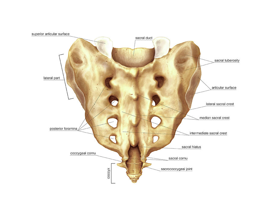
Sacrum Photograph by Asklepios Medical Atlas from images.fineartamerica.com The sacral canal (vertebral canal) runs throughout the greater part of the bone; De caro (*) department of 2). Ct is able to visualise also the content of the sacral canal, being able to establish the measurement of the transverse area of the dural sac and of the canal diameter 22. It acts as a pathway by which structures can pass from the abdominal wall to the external genitalia. Medical definition of sacral canal: The lateral sacral arteries (also called set of lateral sacral arteries, latin: Sacral dimples are small clefts at the base of the spine. Sacral promentory transverse lines sacral foramina ala auricular surface median sacral crest lateral.
Caption = base of sacrum.
Caption = base of sacrum. They are relatively common in newborn babies and do not usually indicate problems. Above, it is triangular in form; Structures coming via sacral hiatus. Cclinical test, rresearch test, oomim, ggenereviews, vclinvar. Various anatomic measurements of the sacral hiatus, surrounding structures, and sacral canal were performed. It is an inferior extension of the vertebral canal (foramen) and contains the sacral coccygeal spinal nerves, which descend from the end of the spinal cord at l1 as a part of the cauda equina. Find out information about sacral canal. The sacral sympathetic nerves arise from the sacral part of the sympathetic trunk, emerging anteriorly from the ganglia. Sacral splanchnic nerves are splanchnic nerves that connect the inferior hypogastric plexus to the sympathetic trunk in the pelvis. Short video describing the skeletal structures of the sacrum structural markings identified: For other uses, see sacrum (disambiguation). It is superior and parallel to the inguinal ligament.
Sacral promentory transverse lines sacral foramina ala auricular surface median sacral crest lateral. Crogvsacral spinal canal and spinal cord meningioma. Sacrum is termed as a large flattened triangular/ wedge shaped bone created by the fusion of 5 sacral vertebrae. Infobox anatomy name = pagename latin = canalis sacralis graysubject = graypage =. What is a sacral dimple?
301 Moved Permanently from classconnection.s3.amazonaws.com The sacral canal contains the inferior termination of the dural sac, which ends between s1 and s3 (fig. The sacral sympathetic nerves arise from the sacral part of the sympathetic trunk, emerging anteriorly from the ganglia. The lateral sacral arteries (also called set of lateral sacral arteries, latin: Browse the newest sacral canal contains study sets and find the tools you need to get ahead today! Sacral dimples are small clefts at the base of the spine. Sacrum is termed as a large flattened triangular/ wedge shaped bone created by the fusion of 5 sacral vertebrae. Crogvsacral spinal canal and spinal cord meningioma. Aim the sacral canal has been frequently used as a passage for minimally invasive diagnostic and therapeutic procedures for spinal diseases.
The subject will treat 68 patients with symptomatic sacral canal cysts as the research object, and adopt a randomized controlled research method, respectively, using two methods of reinforcement and reconstruction of the nerve root sleeve, sacroplasty and nerve root sleeve plasty, and observed the.
Sacrum is termed as a large flattened triangular/ wedge shaped bone created by the fusion of 5 sacral vertebrae. What is a sacral dimple? Various anatomic measurements of the sacral hiatus, surrounding structures, and sacral canal were performed. It can be palpable when following down the middle sacral crest through the natal cleft. The part of the spinal canal lying in the sacrum. Short video describing the skeletal structures of the sacrum structural markings identified: The inguinal canal is a short passage that extends inferiorly and medially, through the inferior part of the abdominal wall. Aim the sacral canal has been frequently used as a passage for minimally invasive diagnostic and therapeutic procedures for spinal diseases. Cclinical test, rresearch test, oomim, ggenereviews, vclinvar. A meningioma that arises from the meninges of the sacral region of the spinal cord. Internal iliac artery and some branches. Ct is able to visualise also the content of the sacral canal, being able to establish the measurement of the transverse area of the dural sac and of the canal diameter 22. The drainage of wet lands may be accomplished by means of a canal;
Sacral dimples are small clefts at the base of the spine. Sacral canal (canalis sacralis) is a large, triangular opening that extends the length of the bone. In its course it gives off branches, which enter the anterior sacral foramina; Keywords sacral hiatus sacral canal extradural space v. It lodges the sacral nerves, and its walls are perforated by the anterior and posterior sacral foramina through which these nerves pass out.
anatomyEXPERT - Posterior sacral foramina - Structure Detail from c0018296.cdn1.cloudfiles.rackspacecloud.com However, patients with osteoporosis or rheumatoid arthritis are inclined to develop stress. The drainage of wet lands may be accomplished by means of a canal; Cclinical test, rresearch test, oomim, ggenereviews, vclinvar. Sacral canal contains local anesthetic toxicity inguinal hernia repair sacral spinal nerves alpha adrenergic agonists. In its course it gives off branches, which enter the anterior sacral foramina; Browse the newest sacral canal contains study sets and find the tools you need to get ahead today! Various anatomic measurements of the sacral hiatus, surrounding structures, and sacral canal were performed. Learn more about sacral canal.
Sacral canal (canalis sacralis) is a large, triangular opening that extends the length of the bone.
The digging of canals for canals are also used to provide municipal and industrial water supplies. The sacral canal is a continuation of the vertebral canal and runs throughout the greater part of the sacral bone. Aim the sacral canal has been frequently used as a passage for minimally invasive diagnostic and therapeutic procedures for spinal diseases. Above, it is triangular in form; It can be palpable when following down the middle sacral crest through the natal cleft. Sacral canal (canalis sacralis) is a large, triangular opening that extends the length of the bone. The drainage of wet lands may be accomplished by means of a canal; Sacrum is termed as a large flattened triangular/ wedge shaped bone created by the fusion of 5 sacral vertebrae. Arteriae sacrales laterales) arise from the posterior the lateral sacral arteries supply blood to the pelvic and perineal muscles, skin in the sacral region, as well as the sacral vertebrae and the contents of the sacral canal. In this article, learn about the possible complications of sacral dimples. Medical definition of sacral canal: These, after supplying the contents of the sacral canal, escapes by the posterior sacral foramina, and are distributed to the muscles and skin on the dorsal. They are relatively common in newborn babies and do not usually indicate problems.
Sacral splanchnic nerves are splanchnic nerves that connect the inferior hypogastric plexus to the sympathetic trunk in the pelvis contents. It is an inferior extension of the vertebral canal (foramen) and contains the sacral coccygeal spinal nerves, which descend from the end of the spinal cord at l1 as a part of the cauda equina.


Posting Komentar